Precision isolation: identifying bacteraemia in the early stages of sepsis
Fast treatment decisions are critical to early intervention in sepsis care, but blood cultures traditionally used to enable diagnosis take time. Dr Nick Collier and Dr Karen Yu consider approaches that could facilitate quicker detection and characterisation of bacteraemia by concentrating bacteria from whole blood.
Advancements in blood culture technology aid the global fight against sepsis, but culture-based diagnostics can still take days. What if bacteraemia could be detected in whole blood instead, with bacteria cells isolated for pathogen identification and Antimicrobial Susceptibility Testing (AST)?
This would be a major step forward, enabling quicker selection of appropriate antibiotics for a condition where every hour of delay increases risk of death.
It’s a challenging goal. After all, sepsis diagnosis requires the isolation of bacteria cells which are outnumbered billions-to-one by other cells in the blood. Nevertheless, technologies exist which make it feasible to work with these low concentrations. Strategic selection and integration of the right approaches could make it commercially viable too.
Cost per test is a very important consideration in sepsis diagnostics. Over the years, there have been many exciting technologies that have struggled to achieve a low enough cost to displace the incumbent technologies for routine testing, often being reserved for high-risk cases. This challenge is even greater for whole blood testing without culture, where typically fewer than 10% of samples test positive, although this depends on the clinical setting e.g. ER vs non-ER. This means the cost per test must be lower than that of systems that operate on positive blood cultures.
Isolating bacteria cells in whole blood
Solutions which identify bacteraemia in whole blood using the molecular techniques of Nucleic Acid Amplification Tests (NAAT) and Next-Generation Sequencing (NGS) are attracting significant innovation and investment. Back in 2012, Sagentia Medical supported T2 Biosystems’ development of its T2Dx Polymerase Chain Reaction (PCR) solution. This went on to achieve FDA clearance and has served as a platform for further iterations. Today, diagnostics companies Deepull and Day Zero are working on solutions based on PCR and NGS respectively. Their diagnostic tests benefit from much larger bacterial panels and the ability to detect Antimicrobial Resistance (AMR) based on known molecular resistance markers.
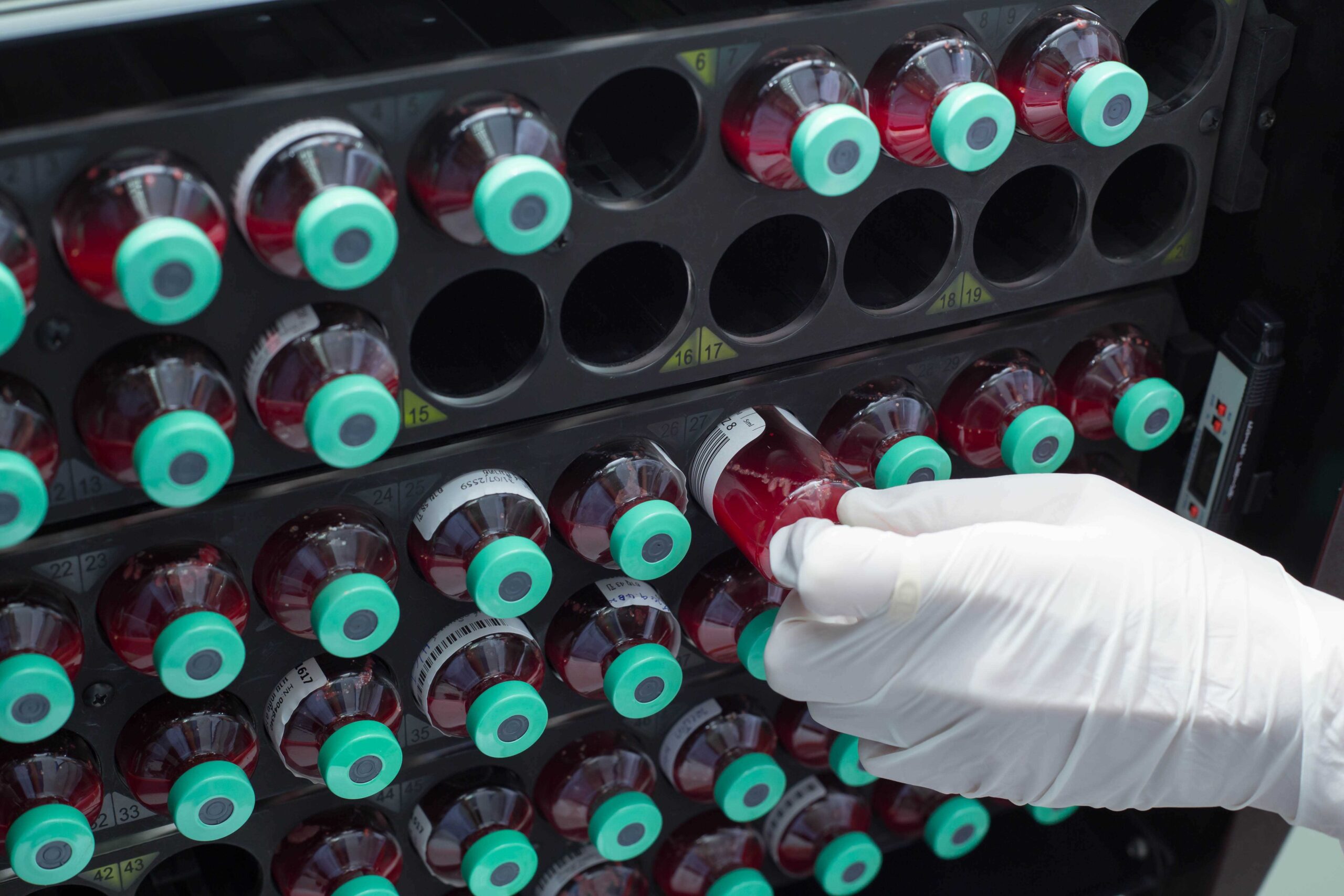
Figure 1: Blood culture bottles in an incubator, where bacterial growth is typically detected within 24–48 hours.
Benefits of NAAT and NGS include the ability to analyse cell free DNA (cfDNA) from non-viable bacteria, which offers the potential of high sensitivity from smaller sample volumes. Yet the many components of whole blood make it a difficult medium for Nucleic Acid (NA) analysis, and the low concentration of bacteria in positive samples exacerbates this. Thus, NA purification and extraction is essential for these techniques. Despite the higher sensitivity, there remains a need for relatively large sample volumes due to the low concentrations of bacterial NA. This comes at a cost; in our experience NA isolation reagents can cost as much as amplification and detection reagents.
So, while the ability of genotypic approaches to identify pathogens and AMR genes offers exciting potential for detecting bacteria in whole blood and providing valuable clinical insights, there are constraints. What’s more, the current standard of care for sepsis diagnosis and treatment decisions often still relies on phenotypic AST. Genotypic AST has several limitations. For one thing, it classifies resistance based on the presence or absence of specific resistance genes, which is only an approximation to actual susceptibility. Whole genome sequencing, while comprehensive, is currently uneconomic and not quick enough for clinical decision-making within the necessary timeframe. The relationship between genotype and susceptibility is continuously evolving too.
In short, phenotypic AST is still required, which necessitates the collection of viable bacteria. This is another area that is fertile for innovation, especially in terms of physical separation techniques for the isolation of any bacteria present in whole blood samples. Separated bacteria can be used in the pathogen identification and AST stages of sepsis diagnosis. This is significantly faster than culturing blood; a recent example being the Momentum Bioscience SepsiSTAT system which isolates bacteria for PCR and phenotypic AST in a matter of hours.
Physical separation techniques
Several methods can be applied to the separation of bacteria from whole blood for sepsis diagnosis. Each brings its own considerations, as the following examples illustrate:
Centrifugation
This versatile separation technique leverages differences in cell density and/or size to achieve isolation. In terms of bacterial recovery, density separation and dilution methods can be very effective. Density separation media can achieve distinct separation of cell types, avoiding the loss of bacteria in the Red Blood Cell (RBC) layer. High dilutions can also result in a high level of bacteria cell recovery, but further concentration steps are then required before pathogen identification and AST are performed.
A recent study (Automatic Culture-free Detection of Bacteria from Blood for Rapid Sepsis Diagnostics) reported impressive bacteria recovery rates of 65% to 95% using centrifugation. Researchers layered a 3mL blood sample on top of a density medium and centrifuged at moderate force (600g) for five minutes. The process also removed >99% of RBCs and 66% of platelets. A subsequent selective lysis step removed the remaining RBCs before concentrating the bacteria by centrifugation for 13 minutes at 1000g.
Microfluidics
The microfluidic method of spiral separation is another effective technique which separates cells based on their size, density, or shape. It’s an inertial approach that uses fluid dynamics to separate and concentrate particles of interest from the sample. In this context, whole blood passes through a spiral channel, creating forces which push particles to various flow positions according to their physical characteristics. Different particle types, such as bacterial cells or blood cells, exit the spiral at different outlets. This allows the target cells to be isolated and recovered for testing.
While spiral separation is very effective, rapid processing of the large sample volumes associated with sepsis diagnosis is a challenge. A study published in 2020 (High resolution and high throughput bacteria separation from blood using elasto-inertial microfluidics) reported that it took 40 minutes to process 1mL of blood using a single spiral at a separation efficiency of 82%. Achieving 90% efficiency took three hours. Parallelisation could be used to overcome this challenge, with multiple spirals running simultaneously so more blood can be processed in less time.
Affinity capture
Particle capture is a highly targeted technique which can be useful to support the diagnosis of bacteraemia. Functionalised particles – such as bubbles or magnetic beads – are used to capture bacteria cells from the whole blood sample. Particle surfaces are coated with affinity molecules (such as antibodies or lectins) which selectively bind and isolate target cells. Momentum Bioscience uses magnetic beads in its SepsiSTAT system mentioned above.
In terms of sepsis diagnosis, many particles are needed due to the low concentration of bacteria cells in positive samples. This makes it difficult to locate active particles which have captured bacteria. Digital detection methods can help but may not be feasible within target test times. Therefore, this technique is often used in combination with others that remove the majority of interfering cells before affinity capture is used as the cleanup step.
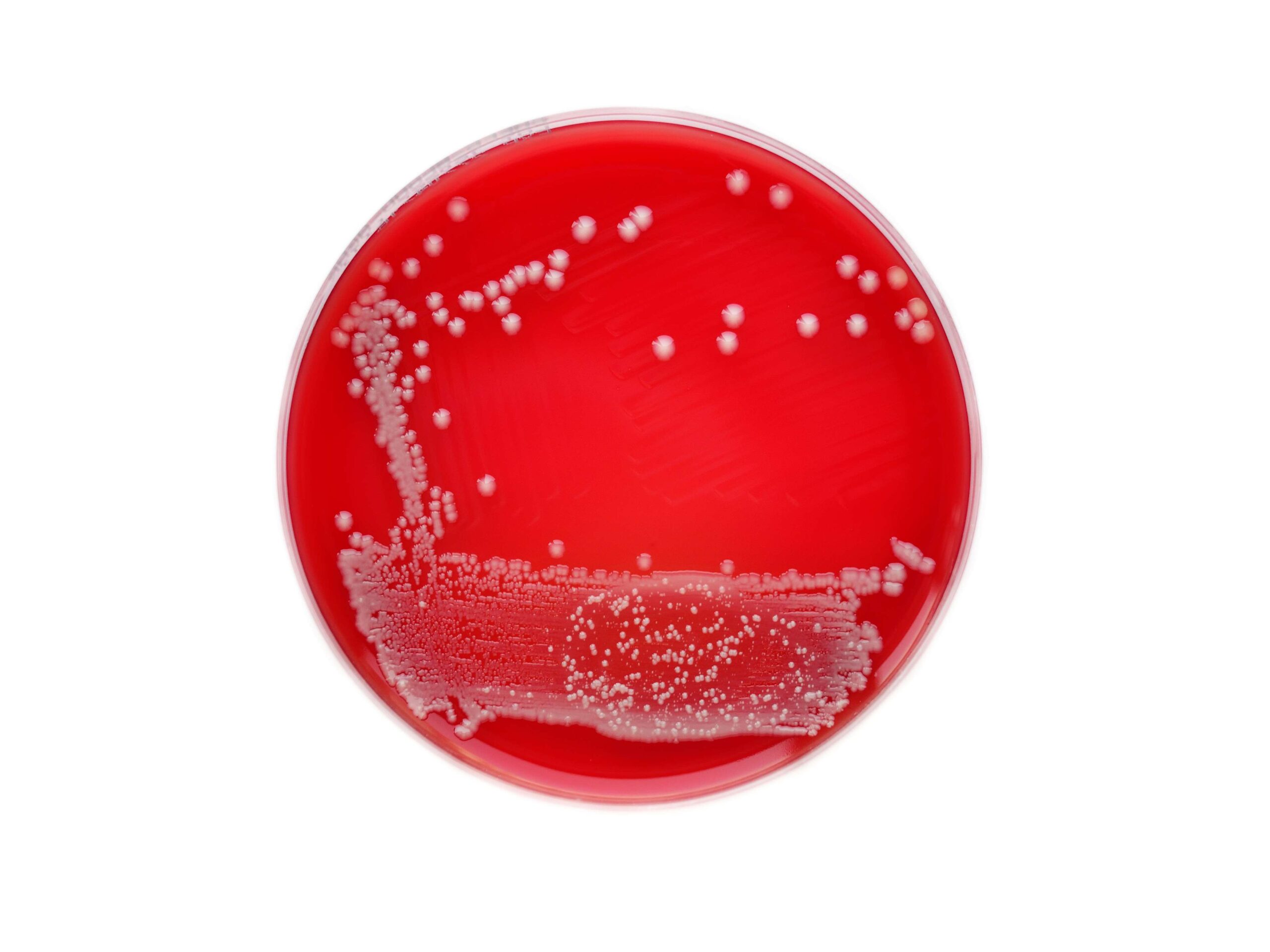
Figure 2: Bacterial colonies growing on an agar plate, typically requiring an additional 18–24 hours before antimicrobial susceptibility testing (AST) can be performed.
Size separation
Since bacteria cells are significantly smaller than RBCs and white blood cells, filtration of whole blood is another feasible option. This simple, passive isolation technique can be very effective, especially when used in combination with other approaches. One noteworthy benefit is that the bacteria can be localised on a planar surface ready for analysis, e.g. using track-etched membranes. Nevertheless, there are limiting factors to consider. These include speed, the clogging of filters, and the risk of incomplete recovery if bacteria cells stick to blood cells or the filter.
Accelerating sepsis diagnosis and more
Clinical evidence suggests that sepsis therapy modification is often delayed while doctors wait for AST results. Since every hour without targeted antibiotic treatment dramatically increases the risk, speed is of the essence.
Developments in NAAT and NGS hold great promise. But constraints surrounding genotypic AMR as the sole basis for guiding clinical decisions mean there is scope for wider innovation.
We believe that innovating around the replacement of blood culture with physical separation techniques will prove highly beneficial. It could fast-track the pre-analytical phase of the existing diagnostic workflow, allowing phenotypic AST to be performed earlier and saving lives.
Physical separation techniques also offer advantages for other diagnostic applications where specific cells must be isolated from a complex cellular background. These include the detection of tumour cells in blood, stool, and other samples. One recent paper (A Cascaded Chip for the High-Purity Capture and Distinguishing Detection of Phenotypic Circulating Tumor Cells in Colon Cancer) explores the use of on-chip spiral separation to isolate circulating tumour cells from whole blood for the diagnosis of colon cancer. Another example that recently received FDA approval is ColoSense from Geneoscopy which detects ribonucleic acid from tumour cells in stools. In this application, it is necessary to isolate the human cells from the very large background of bacterial cells.
How Sagentia Medical can help
Our science, technology, and engineering consultants have a strong track record developing sample preparation techniques to facilitate quick, reliable, and cost-effective medical diagnosis. With expertise spanning biochemistry, microfluidics, system engineering, and more we’re the ideal innovation partner for pre-analytical devices. Contact us if you’d like to arrange an informal discussion about your diagnostic innovation projects.
Read more of our insights

June 20, 2025
Closing the Alzheimer’s gender gap: innovating around early diagnosis and treatment

April 24, 2025
Overcoming technical barriers to blood-based diagnosis of neurodegenerative disease

February 13, 2025
Five critical factors for the commercialisation of women’s health technology